Dissection Master XR Présentation
The app brings high-resolution, three-dimensional datasets of professionally dissected human bodies into augmented reality and provides a high quality learning resource for medical students.
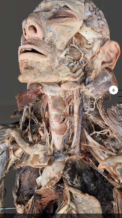
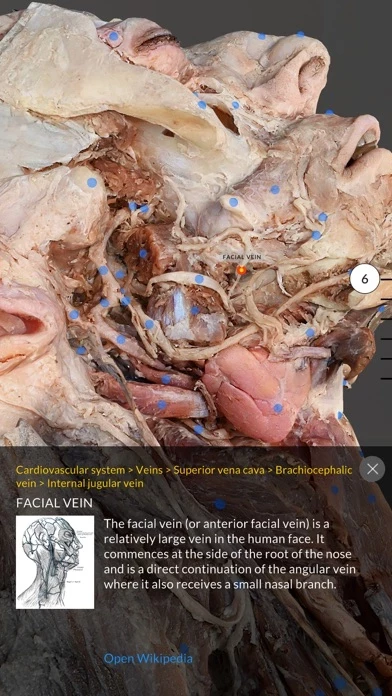
The super high resolution human anatomy models provide visualization of sub-mm structures such as small nerve branches, small blood vessels, lymphatic vessels, and membranes.
Captures d'écran officielles
Détails du produit et description de
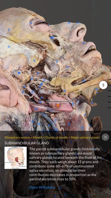
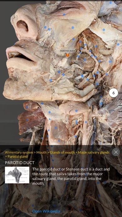
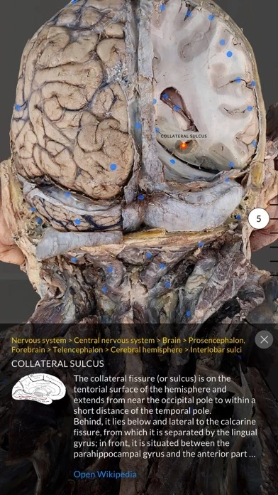
Dissection Master XR is the anatomy lab in augmented reality. The app brings high-resolution, three-dimensional datasets of professionally dissected human bodies into augmented reality and provides a high quality learning resource for medical students. The 3D datasets are displayed with high level of detail and organized in layered groups. Browse from outer muscles to inner organs and study human anatomy with ultimate precision. All datasets are professionally dissected with focus on medical studies and then digitized in very high resolution. Organs and anatomical structures are carefully named and linked to Wikipedia for further information. The app comes with 7 high resolution 3D datasets of head and thorax. Additional datasets will be continuously added. The serial cadaver dissections are designed to teach 3-dimensional relationships normally visible only during a dissection or an operating room procedure. The super high resolution human anatomy models provide visualization of sub-mm structures such as small nerve branches, small blood vessels, lymphatic vessels, and membranes. Included Datasets: Layer 1 · superficial head and neck · superficial fascia and muscles of pectoral region · arms and back Layer 2 Continuation of Model 1 dissection plus: · parotid, buccal regions · anterior triangle of the neck · superficial posterior brain and dura mater · superficial and intermediate muscles of the pectoral region, back, and shoulder · cross section of abdomen at T12 Layer 3 Continuation of Model 2 dissection plus: · anterior and posterior triangles of the neck · temporal region · inferior orbit and infraorbital region · body of mandible (external) · suboccipital triangle, deep back, deep shoulder · superficial mediastinum · superficial posterior brain Layer 4 Continuation of Model 3 dissection plus: · maxillary sinus · infratemporal region · superficial central mediastinum · deep muscles of superior back · deep anterior and posterior triangles of neck Layer 5 Continuation of Model 4 dissection plus: · left anterior descending artery and right-ventricle · cervical ventral rami · infratemporal fossa · frontal sinus · thoracic laminectomy and spinal cord · dural venous sinuses · coronal section of parietal/temporal lobes of brain Layer 6 Continuation of Model 5 dissection plus: · cervical spinal cord and deep suboccipital region · deep shoulder · right coronary artery and right heart chambers and valves · falx cerebri and cross section of supraventricular frontal/parietal lobes · deep face and infratemporal fossa · deep neck · axillary region · superior mediastinum · brachial plexus Layer 7 Continuation of Model 7 dissection plus: · face hemisection (oral and nasal cavities) · orbit · cranial nerves · brainstem · basal nuclei · complete brachial plexus · posterior mediastinum · left heart chambers and valves · rotator cuff; inferior diaphragm; larynx Layer 8: coming soon